Scanning Electron Microscope images
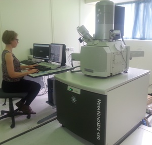
 Materials engineer Dr Ruth Knibbe uses a scanning electron microscope (SEM) to image her own samples and samples for other scientists.
Unlike a light microscope, an SEM uses a focused beam of electrons to produce images from the top surface of a sample, with every location giving a slightly different signal.
In an interview for Our Changing World, Ruth Knibbe explains that different scientists use an SEM for different purposes. “[Biologists] would use this microscope because you can get that high detailed topography information,” Another big benefit of a scanning electron microscope for all types of scientists, as compared with a light microscope, is the huge depth of field which gives SEM images their characteristic, almost 3D-like, quality.
Here are some SEM images of a Bee. The bee died of natural causes and is coated with gold/palladium to provide electrical conductivity to the surface.
All SEM photos courtesy Ruth Knibbe.
The images in this gallery are used with permission and are subject to copyright conditions.


