Dr Melanie McConnell.
Photo: Supplied.
There's a revolution going on in cell biology and one of the people leading it is Dr Melanie McConnell from Victoria University of Wellington.
She was part of a group that made a very unusual discovery. About five years ago the team saw mitochondria (the structure that creates the energy to power a cell) being shuffled between cells; a world first.
This transfer of mitochondria between cells creates opportunities to develop new ways of fighting diseases like Parkinson's, Alzhheimer's and cancer.
She has just been awarded a NZ$1 million grant from the Health Research Council of New Zealand to further research how injured cells can enlist the help of healthy cells living nearby.
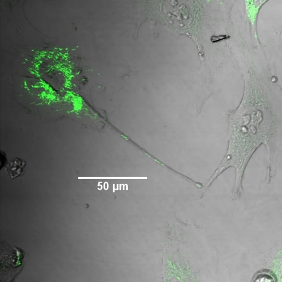
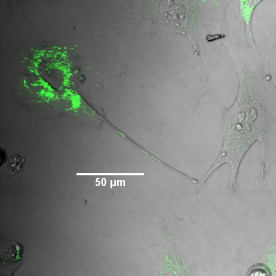
Human astrocytes - one of the most frequent brain cells - growing in a dish in the lab. The mitochondria in the cell on the upper le; have been labeled with a fluorescent green dye. An ‘arm’ containing mitochondria has extended from the labeled cell, and is in contact with an adjacent cell. The line indicates 50 microns, or 1/20th of a millimetre. Image by Remy Schneider, VUW/MIMR
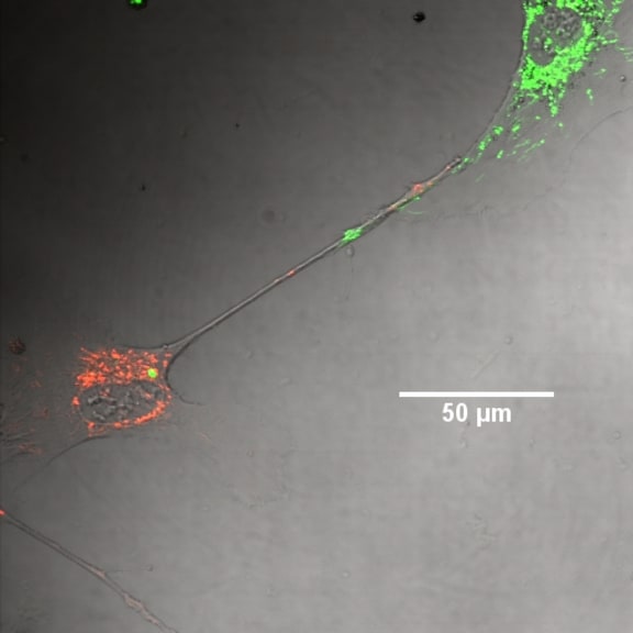
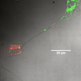
Human astrocyte cells in culture, with mitochondria labeled either red or green. A two-way connection between cells has been made, with green mitochondria entering the ‘red’ cell, and red mitochondria entering the ‘green’ cell. Scale bar represents 50 microns, or 1/20th of a millimetre. Image by Remy Schneider, VUW/MIMR.
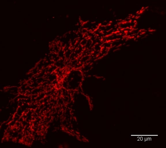
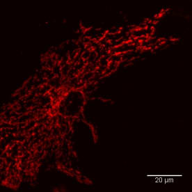
High resolution image of a mouse astrocyte cell grown in culture, with mitochondria stained in red. The scale bar represents 20 microns, or 1/50th of a millimetre. Image by Remy Schneider, VUW/MIMR.
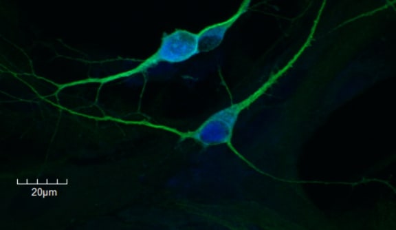
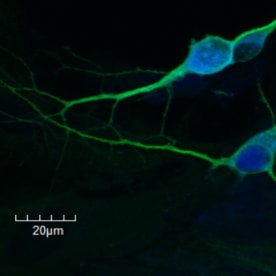
Developing neurons grown in culture, stained with a neural-specific protein in green (MAP2). The nucleus of the cell is stained blue. The scale bar represents 20 microns, or 1/50th of a millimetre. Image by Remy Schneider, VUW/MIMR

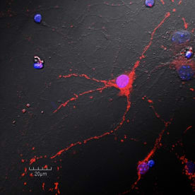
Developing neurons grown in culture, with mitochondria stained in red. The nucleus of the cell is stained blue/ purple. The scale bar represents 20 microns, or 1/50th of a millimetre. Image by Remy Schneider, VUW/MIMR.
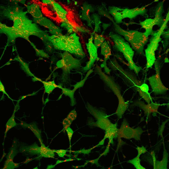

Mixed culture of developing neurons and astrocytes in culture (green) with low level of mitochondrial staining, red.