Navigation for This Way Up
Mitochondria
Mitochondria (the structure that creates the energy to power a cell) being shuffled between cells.
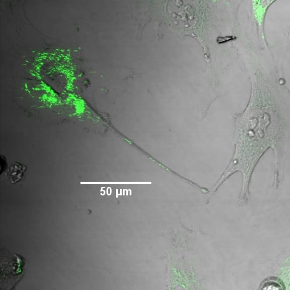

Human astrocytes - one of the most frequent brain cells - growing in a dish in the lab. The mitochondria in the cell on the upper le; have been labeled with a fluorescent green dye. An ‘arm’ containing mitochondria has extended from the labeled cell, and is in contact with an adjacent cell. The line indicates 50 microns, or 1/20th of a millimetre. Image by Remy Schneider, VUW/MIMR
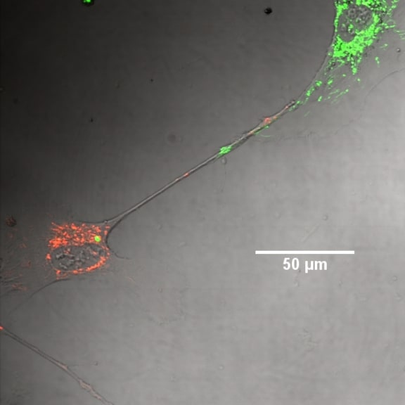

Human astrocyte cells in culture, with mitochondria labeled either red or green. A two-way connection between cells has been made, with green mitochondria entering the ‘red’ cell, and red mitochondria entering the ‘green’ cell. Scale bar represents 50 microns, or 1/20th of a millimetre. Image by Remy Schneider, VUW/MIMR.
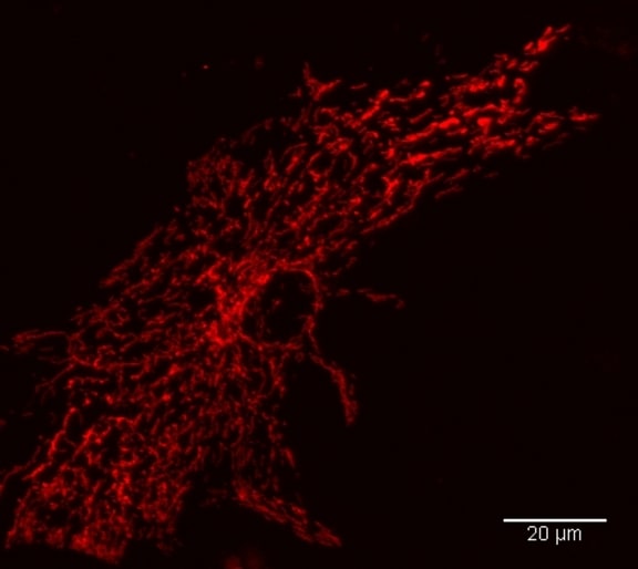

High resolution image of a mouse astrocyte cell grown in culture, with mitochondria stained in red. The scale bar represents 20 microns, or 1/50th of a millimetre. Image by Remy Schneider, VUW/MIMR.
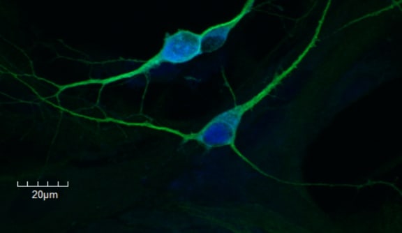

Developing neurons grown in culture, stained with a neural-specific protein in green (MAP2). The nucleus of the cell is stained blue. The scale bar represents 20 microns, or 1/50th of a millimetre. Image by Remy Schneider, VUW/MIMR
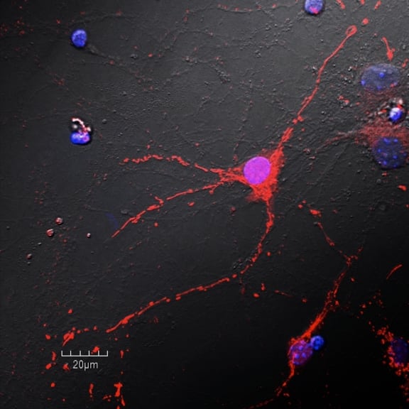

Developing neurons grown in culture, with mitochondria stained in red. The nucleus of the cell is stained blue/ purple. The scale bar represents 20 microns, or 1/50th of a millimetre. Image by Remy Schneider, VUW/MIMR.
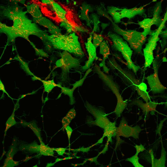

Mixed culture of developing neurons and astrocytes in culture (green) with low level of mitochondrial staining, red.
The images in this gallery are used with permission and are subject to copyright conditions.